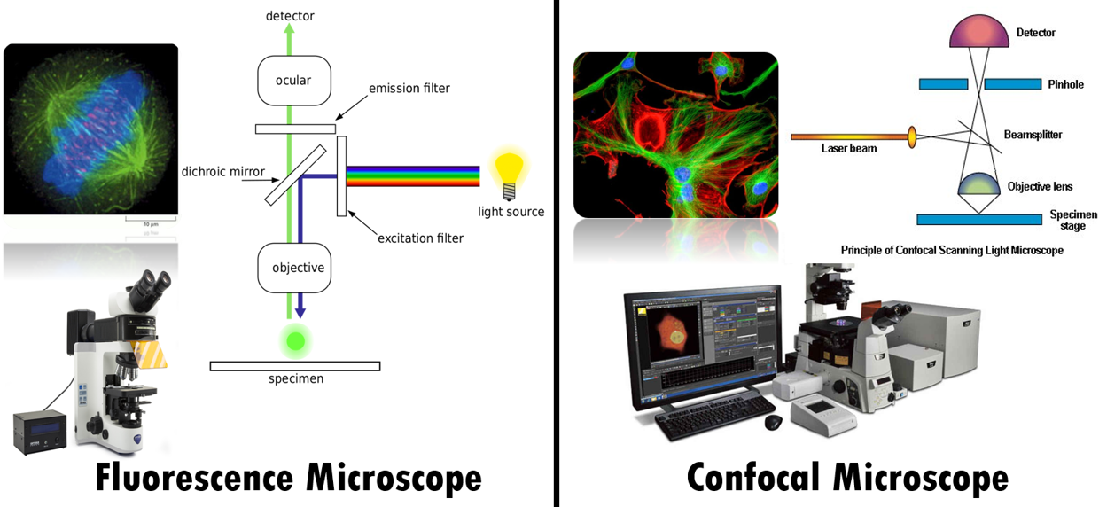
Fluorescence Microscope and Confocal Microscope Comparison
First let us begin with the similarities. Both Fluorescence and Confocal Microscope uses use fluorescent molecules to image specimens.
In
fluorescence microscopy, a specimen is illuminated with a light source that
excites fluorescent molecules in the specimen. The emitted light from these
molecules is then detected and used to create an image.
In
confocal microscopy, point illumination source or a focused laser beam is used
to illuminate the specimen and the emitted light is passed through a pinhole;
to eliminate out-of-focus light. This allows for higher resolution images than
traditional fluorescence microscopy.
Both
can be used to image live cells and thick tissues. Both these microscopes are
widely used in live cell imaging, biomedical research and drug discovery.
|
Fluorescence Microscope |
Confocal Microscope |
|
Principle: Illuminates the entire specimen with a
broad light source and the emitted light from these molecules is then detected
and used to create an image |
A point illumination source is used to scan
the specimen, and the emitted light is passed through a pinhole;
to eliminate out-of-focus light |
|
Lower resolution |
Higher resolution, especially in the z-axis
as out of focus light is eliminated |
|
Deeper depth of field, can image thick
specimens |
Very shallow depth of field that is only
a very thin slice of the specimen is in focus at any given time |
|
Does not use a confocal pinhole. |
Uses a confocal pinhole to reject
out-of-focus light. |
|
Can image multiple structures with a single
fluorescent label (single labeling). |
Requires multiple fluorescent labels
(multiple labeling) to image multiple structures like chromosome, spindle
fibers etc |
|
Acquires images all at once, which is faster. |
Acquires images point-by-point, which can be
time-consuming. |
|
3D imaging possible but not fine as confocal
microscope |
3D imaging possible with high resolution |
|
Imaging of large specimens, such as whole
cells or tissues |
Application:
Imaging of thick tissues, live cells, and single molecules |
|
Can be used for quantitative analysis, but effective
as confocal microscopy. |
Can be used for quantitative analysis like no.
of cells, size of cell structures etc. |
|
Less expensive |
More
expensive |
Image credit: https://www.olympus-lifescience.com/
Watch our simplified video on Types of Microscope
Advantages of confocal microscopes:
- Higher resolution
- Shallow depth of field
- Ability to image thick tissues and live cells
- Good for imaging single molecules
- Less expensive
- Can image thicker specimens
- Good for imaging large specimens
A great way to thank and support us.
- Visit our TPT store Parts of a Microscope Worksheet Free
- Download free resources or purchase some.
- Please rate the product and follow us on store.
Thank you so much

Post a Comment
We Love to hear from U :) Leave us a Comment to improve this site
Thanks for Visiting.....